September 13, 2026 · Clinical Pearl · Cardiomyopathy
Dynamic LVOT Obstruction: How Dynamic Is “Dynamic”? One Doppler Image Tells the Story
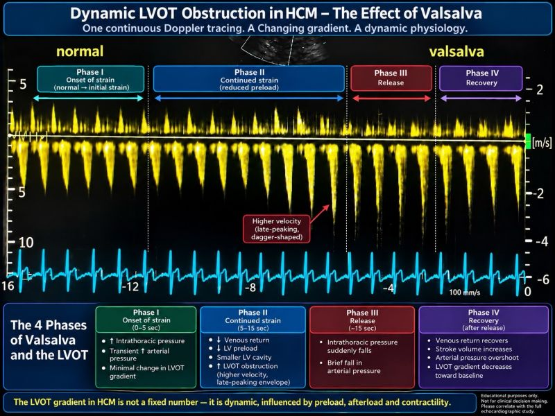
This continuous-wave Doppler tracing beautifully demonstrates why LVOT obstruction in hypertrophic cardiomyopathy is called dynamic. During Valsalva, loading conditions change—and in this single continuous tracing, we can watch the LVOT Doppler envelope change with them.
This CW Doppler tracing beautifully demonstrates why LVOT obstruction in hypertrophic cardiomyopathy is called dynamic.
During Valsalva, loading conditions change; and in this single continuous tracing, we can actually watch the LVOT Doppler envelope change with them.
The 4 phases of Valsalva:
Phase I — Onset of strain: Intrathoracic pressure rises.
Phase II — Continued strain: Venous return falls → LV preload falls → LV cavity becomes smaller.
In obstructive HCM, this can worsen systolic anterior motion and LVOT obstruction. On Doppler, the velocity increases and the envelope becomes more pronounced and late-peaking—the classic “dagger-shaped” profile.
Phase III — Release: Intrathoracic pressure suddenly falls.
Phase IV — Recovery: Venous return and stroke volume recover.
What makes this tracing particularly educational is seeing the changing height and curvature of the Doppler envelopes within one continuous image.
It captures the physiology in real time: Changing preload → changing LV geometry → changing LVOT obstruction.
The LVOT gradient in HCM is not simply a number measured on an echo. It is a dynamic physiologic phenomenon.
#HCM #HypertrophicCardiomyopathy #Echocardiography #CardiacImaging #Cardiology #HeartFailure
This is for informational purposes only. For medical advice or diagnosis, consult a professional.
Important notice
This content is for educational purposes only and is not medical advice for any individual patient. Patients should discuss diagnosis and treatment decisions with their own physician, or request a clinical consultation.