July 1, 2026 · Clinical Pearl · Cardiomyopathy
Beyond the Ejection Fraction: The Role of Cardiac MRI in Heart Failure
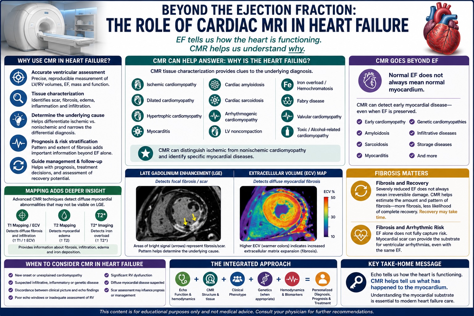
For decades, much of heart failure imaging has revolved around a single number: the ejection fraction. But EF tells us how the heart is functioning—it does not necessarily tell us why it is failing. This is where cardiac MRI has changed the evaluation of heart failure.
For decades, much of heart failure imaging has revolved around a single number: the ejection fraction (EF).
Is the EF 25%? 40%? 55%?
That number remains important. It helps classify heart failure and guides therapies ranging from guideline-directed medical therapy (GDMT) to ICD and CRT consideration. But EF tells us how the heart is functioning—it does not necessarily tell us why it is failing.
This is where cardiac magnetic resonance imaging (CMR) has changed the evaluation of heart failure.
Modern CMR allows us to move beyond simply measuring ventricular function and ask a more important question: What is happening inside the myocardium?
Echocardiography remains the starting point
Echocardiography remains the first-line imaging modality for most patients with heart failure. It is widely available, inexpensive, repeatable, and provides tremendous information about ventricular function, valves, filling pressures, pulmonary pressures, and hemodynamics.
CMR should not replace echocardiography. Instead, it should complement echo when knowing the underlying myocardial substrate could change diagnosis, prognosis, or treatment.
The AHA/ACC/HFSA heart failure guideline recognizes CMR as useful in selected patients with heart failure or cardiomyopathy, particularly when additional information is needed regarding cardiac structure, function, or the cause of myocardial dysfunction.
1. CMR provides extremely accurate ventricular assessment
CMR provides highly reproducible measurements of LV and RV volumes, ejection fraction, ventricular mass, wall thickness, regional wall motion, and chamber morphology.
This can be especially valuable when echocardiographic windows are limited or when accurate assessment of the right ventricle is important.
But measuring EF may actually be the least interesting part of a heart failure MRI.
The real power of CMR is tissue characterization.
2. Late gadolinium enhancement: looking for myocardial scar
Late gadolinium enhancement (LGE) allows us to identify areas of myocardial fibrosis and injury.
More importantly, the pattern of fibrosis can provide clues to the underlying disease.
For example:
Subendocardial or transmural LGE → Think ischemic injury / prior myocardial infarction.
Mid-wall fibrosis → Often associated with dilated cardiomyopathy.
Subepicardial or patchy LGE → May suggest myocarditis or inflammatory cardiomyopathy.
Characteristic infiltrative patterns → Can raise suspicion for diseases such as cardiac amyloidosis or sarcoidosis.
The distribution of scar can therefore help distinguish ischemic from nonischemic cardiomyopathy and narrow the differential diagnosis of otherwise unexplained heart failure.
3. CMR can help answer: Why does this patient have heart failure?
Consider the patient presenting with a newly discovered EF of 25%.
The echocardiogram tells us: The ventricle is failing.
CMR may help tell us: Why.
Potential diagnoses that CMR can help identify or support include ischemic cardiomyopathy, dilated cardiomyopathy, myocarditis, cardiac sarcoidosis, cardiac amyloidosis, hypertrophic cardiomyopathy, arrhythmogenic cardiomyopathy, LV noncompaction, Fabry disease, and iron overload/hemochromatosis.
Current cardiomyopathy guidelines place considerable emphasis on this ability to characterize the myocardial phenotype rather than treating every reduced EF as the same disease.
4. MRI can identify disease even when the EF is preserved
This may be one of the most important concepts. Normal EF does not necessarily mean normal myocardium.
A patient can have preserved systolic function while already developing myocardial fibrosis, inflammation, infiltration, or other tissue abnormalities.
This is particularly relevant in early cardiomyopathy, cardiac amyloidosis, sarcoidosis, myocarditis, hypertrophic cardiomyopathy, and genetic cardiomyopathies.
The 2023 ESC cardiomyopathy guidelines specifically incorporated nonischemic ventricular scar and other CMR tissue-characterization abnormalities into the phenotypic description of cardiomyopathy. Scar may sometimes be an important clue to myocardial disease even without major ventricular dilation or systolic dysfunction.
That represents an important change in how we think about cardiomyopathy.
5. Mapping takes CMR beyond visible scar
LGE is powerful, but newer CMR techniques provide even more information.
T1 mapping and extracellular volume (ECV) can help identify diffuse myocardial abnormalities that may not produce a discrete area of LGE.
They can provide information about processes such as fibrosis, infiltration, edema, and myocardial remodeling.
T2-based imaging can help identify myocardial edema and inflammation, while T2* imaging has an important role in assessing myocardial iron deposition.
The combination of cine imaging, LGE, T1/T2 mapping and ECV has turned MRI from primarily an anatomical test into a form of noninvasive myocardial tissue characterization.
6. Fibrosis may tell us something about recovery
This becomes particularly interesting in patients with potentially reversible cardiomyopathies.
Consider alcohol-related cardiomyopathy, tachycardia-mediated cardiomyopathy, myocarditis, and some forms of toxic cardiomyopathy.
A severely reduced EF does not automatically mean that myocardial function cannot recover.
There is a major biological difference between dysfunctional myocardium and myocardium that has undergone extensive irreversible fibrosis.
CMR can help characterize that substrate.
In practical terms, we are increasingly asking not simply: 'How low is the EF?' but: 'How much myocardial injury and fibrosis are present?'
CMR cannot perfectly predict recovery in an individual patient, but the myocardial substrate adds important information beyond EF alone.
7. Fibrosis also matters for arrhythmic risk
Another emerging concept is that EF alone may not fully capture sudden cardiac death risk.
Two patients can both have an EF of 35% yet have very different myocardial substrates.
One may have relatively little fibrosis. Another may have substantial scar capable of providing the substrate for ventricular arrhythmias.
This is particularly relevant in genetic and nonischemic cardiomyopathies, where scar pattern, genotype, ventricular function, arrhythmia burden, family history, and other clinical variables increasingly contribute to individualized risk assessment.
The ESC cardiomyopathy guidelines emphasize CMR scar assessment as part of diagnosis and risk stratification rather than relying on ventricular function alone.
The future: EF + phenotype + tissue
Heart failure imaging is evolving.
The traditional approach was: Heart failure → Echo → EF → Treatment.
The emerging approach is: Heart failure → Echo → Phenotype → CMR when appropriate → Tissue characterization → Etiology → Risk stratification → Personalized treatment.
Not every heart failure patient needs an MRI.
But in patients with new unexplained cardiomyopathy, suspected infiltrative or inflammatory disease, discordant clinical and echocardiographic findings, poor echo windows, significant RV disease, or a cardiomyopathy in which myocardial scar may influence diagnosis or risk assessment, CMR can provide information that an EF alone simply cannot.
The key message
Echo tells us how the heart is functioning. MRI can help tell us what has happened to the myocardium.
And in modern heart failure care, understanding the myocardial substrate may be just as important as knowing the ejection fraction.
The future of heart failure imaging is therefore not Echo vs. MRI.
It is: Echo + MRI + clinical phenotype + genetics + hemodynamics—all used selectively to understand the disease behind the heart failure.
#CardiacMRI #HeartFailure #Cardiomyopathy #CardiovascularImaging #CMR #EjectionFraction #TissueCharacterization #LGE #Cardiology
Important notice
This content is for educational purposes only and is not medical advice for any individual patient. Patients should discuss diagnosis and treatment decisions with their own physician, or request a clinical consultation.