Perspective · HFpEF
HFpEF: The Diagnosis of (In)Convenience
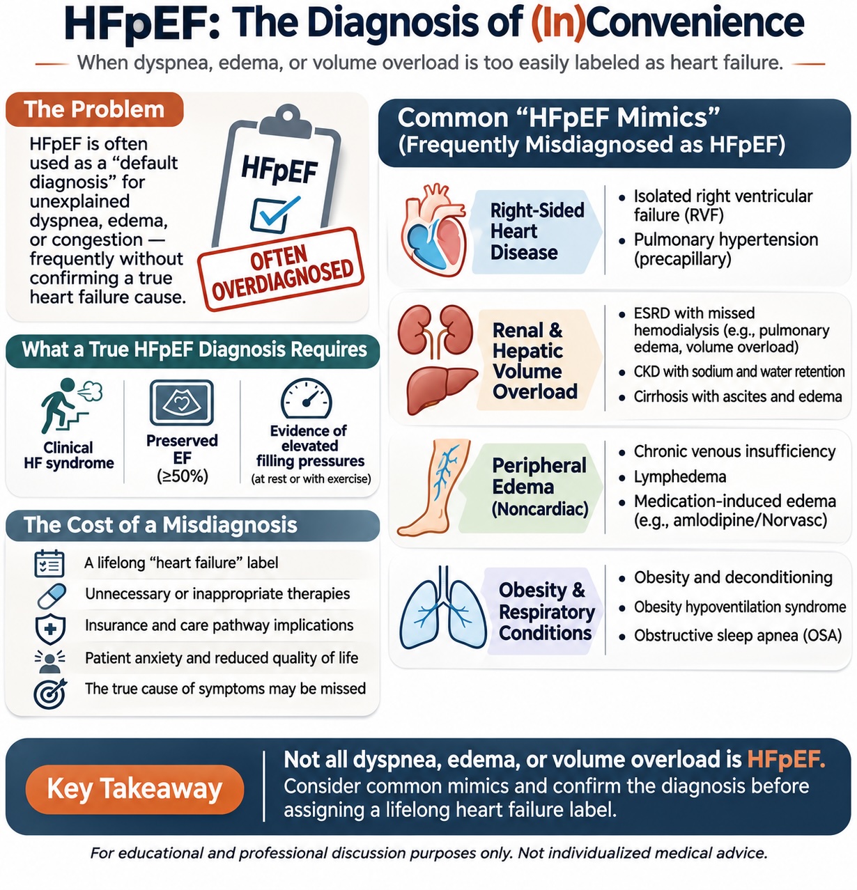
When dyspnea, edema, and volume overload become a lifelong heart failure diagnosis. HFpEF is increasingly used as a convenient explanation for symptoms that may have little to do with intrinsic left ventricular heart failure.
Heart failure with preserved ejection fraction (HFpEF) is increasingly recognized as a major contributor to cardiovascular morbidity and hospitalization. As our understanding of HFpEF has evolved, so have our diagnostic capabilities and therapeutic options.
Yet, alongside this progress, a concerning clinical pattern has emerged: HFpEF is increasingly used as a convenient explanation for symptoms that may have little to do with intrinsic left ventricular heart failure.
A patient presents with shortness of breath, lower-extremity edema, obesity, hypertension, or chronic kidney disease. An echocardiogram demonstrates a left ventricular ejection fraction (LVEF) of 60% and grade I diastolic dysfunction.
The diagnosis? Acute on chronic diastolic congestive heart failure.
But was heart failure ever established? In many cases, the diagnosis is carried forward from one hospitalization to the next, often without reconsideration of its original basis. The problem is that HFpEF has become, at times, a diagnosis of convenience rather than a diagnosis supported by objective evidence.
What Does It Actually Take to Diagnose HFpEF? HFpEF is a clinical syndrome, not simply an echocardiographic finding. Three essential components should guide the diagnosis.
1. A Clinical Heart Failure Syndrome The patient should have symptoms and/or signs consistent with heart failure, including exertional dyspnea, orthopnea, pulmonary congestion, elevated jugular venous pressure, or exercise intolerance. However, these manifestations are not unique to heart failure. Dyspnea, fatigue, and edema occur in numerous cardiac and noncardiac conditions.
2. Preserved Left Ventricular Ejection Fraction HFpEF is generally defined by an LVEF ≥50%. A preserved EF does not establish the diagnosis. It merely identifies the range of ventricular systolic function within which HFpEF may occur.
3. Objective Evidence of Elevated Cardiac Filling Pressures This is often the missing element. Evidence of elevated left-sided filling pressures may be obtained through echocardiographic assessment, natriuretic peptides interpreted in context, or invasive hemodynamic measurements. Importantly, some patients with genuine HFpEF have normal resting filling pressures that become abnormal only during exercise.
Preserved EF + edema + diastolic dysfunction does not automatically equal HFpEF.
The Echocardiogram: Are We Interpreting It Correctly? Echocardiography is central to HFpEF evaluation, but its findings are frequently oversimplified. The presence of grade I diastolic dysfunction is particularly problematic. Impaired myocardial relaxation is common with aging, hypertension, diabetes, and other conditions. It does not necessarily indicate elevated filling pressures or clinical heart failure.
Several echocardiographic parameters provide useful information. The echocardiogram deserves a closer look: • E/e′ >14: supports elevated LV filling pressures. • Septal e′ <7 or lateral e′ <10 cm/s: impaired relaxation. • LAVI >34 mL/m²: suggests chronically elevated filling pressures. • LVH/concentric remodeling: supportive structural findings, but nonspecific.
These are familiar conventional echocardiographic thresholds. Contemporary ASE guidance emphasizes an integrated, multiparametric approach rather than treating individual measurements as definitive.
Left atrial enlargement may result from atrial fibrillation or valvular disease. LV hypertrophy may reflect longstanding hypertension. Reduced e′ velocities can occur without elevated filling pressures. Similarly, patients with HFpEF may have relatively normal resting echocardiographic findings.
The Great HFpEF Mimics: When Everything Becomes Heart Failure One of the most concerning consequences of diagnostic oversimplification is the tendency to label nearly any patient with edema, dyspnea, or volume overload as having HFpEF. Several clinical scenarios deserve particular attention.
1. Isolated Right Ventricular Failure and Pulmonary Hypertension Patients with right ventricular failure frequently present with peripheral edema, elevated jugular venous pressure, ascites, and exercise intolerance. When the LVEF is preserved, these findings may be attributed to HFpEF. However, isolated RV failure from precapillary pulmonary hypertension is fundamentally different from left-sided HFpEF. A patient with severe pulmonary arterial hypertension, RV dysfunction, and normal left-sided filling pressures should not automatically be classified as having HFpEF. The distinction matters because the underlying mechanisms, investigations, and treatment strategies differ substantially.
2. ESRD: The Patient Who Missed Hemodialysis Consider a familiar hospital scenario. A patient with end-stage renal disease misses two hemodialysis sessions and presents with dyspnea, hypertension, pulmonary edema, and peripheral edema. The echocardiogram shows an EF of 60%. The admission diagnosis becomes acute on chronic diastolic CHF exacerbation. But what actually happened? The patient accumulated sodium and water because dialysis was missed. The resulting volume overload may cause pulmonary congestion and elevated cardiac filling pressures without establishing an underlying chronic HFpEF syndrome. After ultrafiltration, the symptoms resolve. Was this truly a heart failure exacerbation, or primarily dialysis-related hypervolemia? This distinction is not merely semantic. It determines whether the patient should carry a lifelong diagnosis of heart failure. Of course, ESRD and HFpEF frequently coexist. The critical question is whether intrinsic cardiac heart failure has actually been demonstrated.
3. Chronic Kidney Disease Without Established Heart Failure Patients with advanced CKD may develop sodium retention, extracellular volume expansion, hypertension, and edema. These patients are often labeled with HFpEF simply because they are volume overloaded and have a preserved EF. Yet renal volume overload and intrinsic cardiac heart failure are not interchangeable diagnoses. The presence of LVH or impaired relaxation does not independently resolve this distinction.
4. Cirrhosis and Hepatic Volume Overload Patients with advanced cirrhosis may develop ascites, peripheral edema, and pleural effusions through portal hypertension, renal sodium retention, and alterations in vascular and oncotic physiology. These findings can closely resemble systemic congestion from heart failure. While cirrhotic cardiomyopathy and HFpEF can coexist, the presence of ascites and edema alone does not establish a cardiac cause. Misclassification may divert attention from the underlying hepatic disease and its appropriate management.
5. Chronic Venous Insufficiency and Lymphedema Bilateral lower-extremity edema is frequently interpreted as evidence of heart failure. Yet chronic venous insufficiency and lymphedema are common causes of persistent peripheral swelling. A patient with chronic leg edema, normal JVP, no pulmonary congestion, and no objective evidence of elevated left-sided filling pressures should not automatically receive an HFpEF diagnosis because an echocardiogram shows preserved EF and impaired relaxation. These patients may receive repeated diuretic prescriptions without addressing the underlying venous or lymphatic pathology.
6. Obesity and Physical Deconditioning Obesity is strongly associated with HFpEF, but it is also an independent cause of exertional dyspnea and reduced exercise capacity. Patients with severe obesity may experience increased respiratory workload, reduced chest wall compliance, impaired exercise tolerance, and substantial deconditioning. A patient with BMI 45, exertional dyspnea, and grade I diastolic dysfunction may have HFpEF—but may also have predominantly obesity-related respiratory limitation. The distinction can be difficult, particularly because obesity may suppress natriuretic peptide concentrations and genuine obesity-related HFpEF can be missed. The answer is not to dismiss HFpEF in obesity, but to avoid assuming it.
7. Obesity Hypoventilation Syndrome and Obstructive Sleep Apnea Obesity hypoventilation syndrome and obstructive sleep apnea can cause fatigue, exertional dyspnea, sleep disruption, hypoxemia, and sometimes pulmonary hypertension or right-sided cardiac dysfunction. These patients frequently have obesity, hypertension, and preserved EF—the same clinical characteristics commonly associated with HFpEF. Yet their symptoms may be primarily respiratory or sleep-related rather than attributable to elevated LV filling pressures. A diagnosis of HFpEF should not replace a careful evaluation for sleep-disordered breathing and hypoventilation.
8. Medication-Induced Edema: The Amlodipine, Nifedipine Example A patient with hypertension begins amlodipine (Norvasc) and subsequently develops bilateral ankle swelling. The echocardiogram demonstrates EF 65% and mild diastolic dysfunction. The patient is labeled with HFpEF and started on diuretics. However, dihydropyridine calcium-channel blockers can cause peripheral edema through changes in precapillary arteriolar tone and capillary hydrostatic pressure. This is not necessarily systemic volume overload or heart failure. Before assigning a chronic cardiac diagnosis, medication review should be a fundamental part of the evaluation.
When the Diagnosis Is Uncertain, Should We Prove It? The challenge is that HFpEF is not always straightforward to diagnose. Some patients have convincing clinical symptoms but inconclusive resting echocardiographic findings. Others have multiple comorbidities that increase the likelihood of HFpEF while simultaneously providing alternative explanations for their symptoms. This is precisely where structured diagnostic tools become valuable.
The H₂FPEF score incorporates six clinical and echocardiographic variables: obesity, antihypertensive medication burden, atrial fibrillation, pulmonary artery systolic pressure, age, and E/e′. The HFA-PEFF algorithm incorporates functional, morphological, and biomarker domains, with additional testing recommended when the probability remains intermediate. Neither tool is perfect, but both encourage a more systematic assessment. Should we routinely incorporate one of these approaches when evaluating patients with suspected HFpEF rather than relying on an isolated echocardiographic phrase?
For patients with persistent diagnostic uncertainty, especially unexplained exertional dyspnea, exercise hemodynamic assessment may be appropriate. Invasive measurements supporting HFpEF include: • Resting pulmonary capillary wedge pressure (PCWP) ≥15 mmHg. • Peak exercise PCWP ≥25 mmHg during supine exercise. • Exercise PCWP/cardiac output slope >2 mmHg/L/min. These measurements require careful interpretation, including consideration of loading conditions, exercise position, respiratory pressure changes, and other causes of elevated filling pressures.
Not every patient needs right-heart catheterization. However, when noninvasive evaluation remains inconclusive and the diagnosis would materially alter treatment or long-term care, more definitive physiological assessment deserves consideration. Perhaps we should be more willing to question a diagnosis than to perpetuate it.
The Hidden Cost of Misdiagnosis: A Lifelong Label The greatest consequence of HFpEF overdiagnosis is not simply inaccurate medical documentation. It is the impact on the patient. Once “heart failure” enters the medical record, it often becomes a permanent fixture. It appears on problem lists, discharge summaries, referral documentation, insurance records, and subsequent clinical encounters. Future clinicians may accept the diagnosis as established without reviewing the original evidence. The consequences can be substantial.
• Unnecessary treatment: patients may receive chronic diuretics or other therapies that are not appropriate for their underlying condition, potentially contributing to hypotension, electrolyte abnormalities, or renal dysfunction. • Diagnostic anchoring: new symptoms may automatically be attributed to heart failure, delaying recognition of pulmonary, renal, hepatic, venous, or other disorders. • Healthcare utilization: an inaccurate diagnosis may influence referrals, investigations, monitoring, and clinical pathways. • Psychological burden: being told that one has chronic heart failure can profoundly affect a patient's perception of their health, prognosis, and future. • Difficulty reversing the diagnosis: even when later investigations fail to support HFpEF, removing the label from the medical record can be surprisingly challenging.
For a patient who simply missed dialysis, developed amlodipine-associated edema, or has chronic venous insufficiency, a lifelong diagnosis of heart failure may represent a significant and avoidable disservice.
HFpEF Is Real. So Is Its Potential for Misdiagnosis. The purpose of this discussion is not to minimize HFpEF. It is a serious clinical syndrome with substantial morbidity, and failure to recognize genuine HFpEF can also delay effective treatment. Nor should the presence of CKD, obesity, pulmonary hypertension, or other comorbidities exclude HFpEF. These conditions frequently coexist.
The objective is diagnostic precision. We should distinguish intrinsic cardiac heart failure from other causes of dyspnea, edema, and volume overload. We should interpret echocardiographic findings within their proper clinical context. We should use validated diagnostic frameworks and pursue further testing when uncertainty remains consequential.
Above all, we should recognize that assigning a diagnosis of heart failure is not a trivial decision. Not all volume overload is heart failure. Not all dyspnea is HFpEF. Not all diastolic dysfunction is diastolic heart failure.
HFpEF should not become a diagnosis of convenience. It should remain a diagnosis supported by thoughtful clinical evaluation and objective evidence. Perhaps one of the most important advances we can make in HFpEF care is ensuring that the patients we diagnose and treat actually have the disease.
Disclaimer: This article is intended for medical education and professional discussion. It does not constitute individualized medical advice. Clinical decisions should be guided by comprehensive patient evaluation, current professional guidelines, and appropriate specialist judgment.
Important notice
This content is for educational purposes only and is not medical advice for any individual patient. Patients should discuss diagnosis and treatment decisions with their own physician, or request a clinical consultation.